calcaneal fractures what the surgeon needs to know
Original Editor - Hajar Abdelhadji, Roxann Musimu, Dylan Van Calck Top Contributors -
Hajar Abd,
Kim Jackson,
Manisha Shrestha,
Admin,
Lisa De Poorter1,
Roxann Mus,
Elien Vanderlinden,
Laura Ritchie,
Dylan Van Calck,
Lucinda hampton,
Patti Cavaleriand
Merlin RoggemanDefinition / Description [edit | edit source]
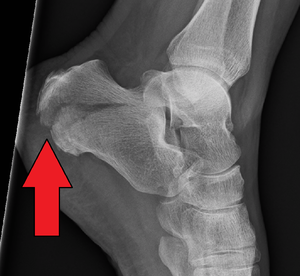
Radiological image of calcaneus fracture( lateral view)
A calcaneus fracture is a heel bone fracture. It is a rare type of fracture but has potentially debilitating results. Traditionally, a burst fracture of the calcaneus was known as"Lovers Fracture" as the injury would occur as a suitor would jump off a lover's balcony (centric loading) to avoid detection.[1]
Clinically Relevant Anatomy [edit | edit source]
A skilful understanding of the anatomy of the calcaneus is essential in determining the patterns of injury and handling goals and options.
Calcaneus is the largest talar bone out of seven tarsal bones which together with the talus form hind-pes. The calcaneus has a relatively thin cortex. It has four facets: 1 anteriorly which articulate with cuboid forming calcaneocuboid joint and three superiorly ( anterior, middle, and posterior, with the posterior facet representing the major weight-bearing surface) which articulate with talus forming talocalcaneal joint (subtalar joint). Subtalar joint allows inversion and eversion of the foot.[2] [1]
The interosseous ligament and medial, lateral, and posterior talocalcaneal ligaments provide additional support for the joint. The sustentaculum tali is a medial bony projection supporting the neck of the talus. The tibial avenue, nerve, posterior tibial tendon, and flexor hallucis longus tendon are located medially to the calcaneus and are at risk for impingement with a calcaneal fracture and, every bit are the peroneal tendons located on the lateral aspect of the calcaneus. This also makes surgical approach challenging. The lateral side of the calcaneus and its apartment nature is highlighted as the most advantageous for internal fixation, just the poor soft tissue encompass challenges wound healing.[3]These anatomic landmarks are important because fractures associated with these areas may cause involve joint involvement, tendon and neurovascular injury.[3]
The calcaneus has four of import functions:
- Acts every bit a foundation and support for the body'due south weight
- Supports the lateral column of the foot and acts as the principal articulation for inversion/eversion
- Acts equally a lever arm for the gastrocnemius muscle complex
- Makes normal walking possible
For more detailed anatomy see Ankle and Foot and Calcaneus
Epidemiology/Etiology [edit | edit source]
- Calcaneal fractures account for 1-ii% of all fractures[iv] and 60% of tarsal fractures[4].
- Less than 10% present as open up fractures.
- Earlier, calcaneum fracture was predominately in male equally they used to practice more than industrial work. Just contempo studies suggest regional variation in male and female predominance.[1]
- 75% of the calcaneus fracture is intra-articular and the prognosis of intra-articular fracture is poor[iv].
- Calcaneal fractures are rare in children.[1]In those viii-14 years-old, threescore% of calcaneal fractures are extra-articular. This number increased to xc% for those under seven years-erstwhile[4].
- 20-25% of the cases with a calcaneal fracture is associated with compression fractures of the lumbar vertebrae. [5]
- Most patients with calcaneus fractures are young, with the 20-39 age group the most common.
- Risk factors for calcaneal fractures include: osteoporosis, diabetes mellitus, peripheral neuropathy, osteomalacia, and long-term immunosuppressive therapy[4].
Mechanism of Injury / Pathological Process [edit | edit source]
- Calcaneal fractures are by and large the result of high energy events leading to axial loading of the os.
- Predominantly, falls from height and automobile accidents (a human foot depressed against an accelerator, brake, or floorboard) are common mechanisms of injury. The talus interim as a wedge causes low and thus flatten, widen, and shorten the calcaneal body.
- Calcaneal fractures can too occur with less severe accidents like an ankle sprain or a stress fracture in runners.
- Jumping onto hard surfaces, blunt or penetrating trauma and twisting/shearing events may also cause calcaneus fracture.[1]
- Mostly, injuries occur in isolation. Almost seen concomitant injuries were lower limb (13.2%) or spinal injuries (6.3%).[6]
- The posterior tibial neurovascular bundle runs along the medial aspect of the calcaneal body and is shielded past the sustentaculum tali thusneurovascular injuries are uncommon with calcaneal fractures.[1]
[7]
Characteristics / Clinical Presentation [edit | edit source]
Initially, a patient may present with an to a higher place mentioned traumatic event with the post-obit clinical features:
- Patients will present with diffuse pain, edema, and ecchymosis at the afflicted fracture site.
- The patient is not likely able to bear weight, walk, and move the foot.
- Swelling in the heel surface area
- Plantar ecchymosis extending through the plantar arch of the foot should raise suspicion significantly.
- There may be associated inability of the Achilles tendon, too raising the suspicion of a calcaneus injury.
- Skin quality around the heel must be evaluated for tenting and/or threatened pare. This is especially important in the setting of Natural language-blazon calcaneus fractures.[i]
- Generalized pain in the heel area that commonly develops slowly (over several days to weeks): typically for stress fractures
- Deformity of the heel or plantar curvation: Secondary to the displacement of the lateral calcaneal border outward, there is a possible widening or broadening of the heel.[half-dozen]
Examination [edit | edit source]
- Palpation: Tenderness over calcaneus while squeezing the heel from both sides.[8] A squeeze examination is positive in stress fracture of the calcaneus.
- A thorough neurovascular test is a must. For which pulse rate of ipsilateral dorsalis pedis or posterior tibial can be compared to the contralateral limb.If there is any suspicion of arterial injury and prompt further investigation with angiography or Doppler scanning can be washed.
- Evaluation of all lower extremity tendon part is also necessary.
- Examination of spine fracture equally an associated fracture of calcaneus from axial loading.[1]
Diagnostic Procedures [edit | edit source]
Radiological test:
X-ray: AP, lateral, and oblique plain films of the human foot and ankle are needed. A harris view may be obtained which demonstrates the calcaneus in an centric orientation.
- Axial - Determines principal fracture line and displays the body, tuberosity, middle and posterior facets.
- Lateral - Determines Bohler bending.
- Oblique/Broden'southward view - Displays the caste of deportation of the master fracture line.[2]
CT scan: It is gold standard for traumatic calcaneal injuries.
Bone scan or MRI: are recommended in stress fracture of the calcaneus.
Some of the reference angle and sign in the radiographic images are:
- Mondor's Sign is a hematoma identified on CT that extends along the sole and is considered pathognomic for calcaneal fracture.
- Bohler's Angle is defined as the angle between two lines drawn on manifestly picture show. The showtime line is between the highest point on the tuberosity and the highest point of posterior facet and the 2nd is the highest point on the anterior process and the highest point on the posterior facet. The normal bending is between 20-40 degrees. Information technology may be depressed on plain radiographs if information technology's calcaneus fracture.
- TheCritical Angle of Gissane is defined as the bending between two lines drawn on plain motion-picture show. The first along the anterior downward slope of the calcaneus and the second along the superior upwardly slope. A normal bending is 130-145 degrees. It may be an increase in calcaneus fracture.[one]
Nomenclature [edit | edit source]
Calcaneal fractures tin can be classified into two general categories.
- Extraarticular fractures: Information technology accounts for 25 % of calcaneal fractures. These typically include avulsion injuries of either the calcaneal tuberosity from the Achilles tendon, the anterior process from the bisect ligament, the sustentaculum tali, calcaneal trunk fracture, stress fracture.
[9]
- Intraarticular Fractures: Fractures involving whatever of the three subtalar articulating surfaces are known as intra-articular fractures and are mutual results of loftier force axial loading: a combination of shearing and compression forces produces both the primary and secondary fracture lines. Information technology accounts for the remaining 75%. The talus acts equally a hammer or wedge compressing the calcaneus at the bending of Gissane causing the fracture. An intra-articular fracture tin can be further classified based on these ii classification organization mentioned beneath:[2]
[ten]
Essex-Lopresti classification: It is based on fracture lines using lateral radiographical images.
- Joint depression type with a unmarried verticle fracture line through the angle of Gissane separating the inductive and posterior portions of the calcaneus.
- Tongue type which has the same verticle fracture line as a depression blazon with some other horizontal fracture line running posteriorly, creating a superior posterior fragment. The tuberosity fragment may and then rotate superiorly.[1]
Sander Classification: It is a widely used classification organisation which uses coronal reconstructed CT images showing posterior facet. The Sanders classification organisation is useful non only in treatment planning but in helping to determine prognosis.[3] at that place are 4 types of fracture based on the number and location of the articular fragments.
- Blazon I fractures are nondisplaced.
- Type 2 fractures (two articular pieces) involve the posterior facet and are subdivided into types A, B, and C, depending on the medial or lateral location of the fracture line (more medial fractures are harder to visualize and reduce intraoperatively).
- Type III fractures (iii articular pieces) include an additional depressed heart fragment and are subdivided into types AB, Air-conditioning, and BC, depending on the position and location of the fracture lines.
- Blazon IV fractures (four or more articular fragments) are highly comminuted.[1] [3]
[xi]
Management/Intervention [edit | edit source]
There is no universal treatment or surgical arroyo to all calcaneal fractures. Treatment of calcaneal fractures depends on the private fracture pathoanatomy, accompanying soft-tissue damage, associated injuries, functional demand, and comorbidities of the patient.[12]
The treatment of calcaneus fracture can exist divided into ii types: operative and non-operative treatment.
Operative Care [edit | edit source]
For the majority of patients with the calcaneus fracture, surgery is the correct form of handling[13]. Intraarticular fractures are often treated operatively. The goal of surgery is to reconstruct the overall shape of the calcaneum and joint surfaces are of utmost importance to obtain a practiced functional upshot.[12] This is possible by performing an open reduction and internal fixation of the fracture. At that place are diverse approaches for performing open up reduction and internal fixation. This procedure decreases the possibility of developing arthritis ( post-traumatic subtalar arthritis) and maximizes the potential for inversion and eversion of the foot.[12]
Extraarticular fractures with a substantial hindfoot varus or valgus deformity (>10°) and those with significant flattening, broadening, or shortening of the heel should also be reduced, preferably via small-scale or percutaneous approaches. A unsafe extraarticular subtype of a tongue-type fracture with astringent deportation of the superior margin of the calcaneal tuberosity ("beak fracture") is a surgical emergency.[12]
Non-Operative Intendance [edit | edit source]
Nonoperative management is preferable when in that location is no impingement of the peroneal tendons and the fracture segments are not displaced (or are displaced less than 2 mm).[12] Nonoperative intendance is also recommended when, despite the presence of a fracture, proper weight-begetting alignment has been adequately maintained and articulating surfaces are not disturbed. Extra-articular fractures are generally treated conservatively. Patients who are over the historic period of 65 years quondam or who take pre-existing health atmospheric condition, such as diabetes, neurovascular insufficiency, noncompliance (e.1000., substance abuse), and severe systemic disorders with immunodeficiency and/or poor overall prognosis are too commonly treated using nonoperative techniques.[12]
RICE protocol for soft tissue injury tin can be followed with immobilization in non- operative direction.
Immobilization: A cast, splint, or brace volition hold the basic in your foot in proper position while they heal. A cast needs to article of clothing for half-dozen to 8 weeks — or maybe longer based on follow up radiographic images. During this time, weight-begetting is strictly prohibited.[fourteen]
Physical Therapy Management [edit | edit source]
Subsequently the surgery, agile range of motion exercises may be practiced with small amounts of motion for all joints of the foot and talocrural joint. These exercises are used to maintain and regain the ankle joint movement. When needed for the involved lower extremity, the patient may continue with elevation, icing and compression. During the therapy, the patient will progress to gradual weight bearing. Patients may find this very difficult and painful. The physiotherapist conducts joint mobilisation to all hypomobile joints.
During the treatment, progressive resisted strengthening of the gastrocnemius muscles is done by weighted exercises, toe-walking, ascending and descending stairs and plyometric exercises. When the fracture is healed, the physiotherapist volition progress the weight bearing in more than stressful situations. This therapy consists of gait instruction and balance practice on different surfaces.
These are some outcome measures that can exist used to measure the functional abilities of the patient to meet the prognosis which can be used during the rehabilitation period.
- Lower Extremity Functional Scale (LEFS)
- Foot and Ankle Power Measure out (FAAM)
Pre-Surgery [edit | edit source]
Initial stability is essential for open reduction internal fixation of intraarticular calcaneal fractures.
Preoperative revalidation consist of:
• Immediate elevation of the affected foot to reduce swelling.
• Compression such as pes pump, intermittent pinch devices, or compression wraps as tolerable.
• Instructions for using wheelchair, bed transfers, or crutch walking.[15] [16]
Postal service-Surgery [edit | edit source]
Both the progression of nonoperative and postoperative direction of calcaneal fractures include traditional immobilization and early move rehabilitation protocols. In fact, the traditional immobilization protocols of nonoperative and postoperative direction are like, and are thereby combined in the progression below. [two] Phases 2 and III of traditional and early motility rehabilitation protocols after nonoperative or postoperative care are comparable likewise and are described together below.[6] [17]
Phase I: Weeks 1-iv [edit | edit source]
Goals: [edit | edit source]
- Control oedema and pain
- Preclude extension of fracture or loss of surgical stabilization
- Minimize loss of part and cardiovascular endurance
Intervention: [edit | edit source]
- Cast with the ankle in neutral and sometimes slight eversion,
- Meridian
- Toe curl and active ankle joint (dorsiflexion and plantarflexion)-encourage to do from the offset post-operative solar day.[18]
- Afterwards 2-4 days, instruct in non-weight bearing ambulation utilizing crutches or walker-crutch walking training
- Instruct in wheelchair utilise with an appropriate sitting schedule to limit time involved extremity spends in dependent-gravity position
- Instruct in comprehensive exercise and cardiovascular plan utilizing upper extremities and uninvolved lower extremity
- Strengthening next joint musculature ( hip and knee)
Phase II: Weeks 5-eight [edit | edit source]
Goals: [edit | edit source]
- Control remaining or residual oedema and hurting
- Prevent re-injury or complication of fracture by progressing weight-bearing safely
- Prevent contracture and regain motion at ankle/foot joints
- Minimize loss of function and cardiovascular endurance
Intervention: [edit | edit source]
- Connected elevation, icing, and compression equally needed for involved lower extremity.
- After 6-8 weeks, instruct in fractional-weight begetting ambulation utilizing crutches or walker
- Initiate vigorous practise and range of motion to regain and maintain motion at all joints: tibiotalar, subtalar, midtarsal, and toe joints, including active range of movement in large amounts of movement and progressive isometric or resisted exercises
- Progress and monitor comprehensive upper extremity and cardiovascular program
Phase III: Weeks 9-12 [edit | edit source]
Goals: [edit | edit source]
- Progress weight-bearing status
- Normal gait on all surfaces
- Restore full range of motion
- Restore total strength
- Permit return to previous work status
Intervention: [edit | edit source]
- After nine-12 weeks, instruct in normal total-weight bearing airing with the advisable assistive device equally needed
- Progress and monitor the subtalar joint'southward ability to adapt for ambulation on all surfaces, including graded and uneven surfaces
- Joint mobilization to all hypomobile joints including: tibiotalar, subtalar, midtarsal, and to toe joints
- Soft tissue mobilization to hypomobile tissues of the gastrocnemius circuitous, plantar fascia, or other appropriate tissues
- Progressive resisted strengthening of gastrocnemius complex through the use of pulleys, weighted practise, toe-walking airing, ascending/descending stairs, skipping or other plyometric exercise, pool exercises, and other climbing activities
- Work hardening programme or activities to permit return to work betwixt 13- 52 weeks.
Implant Removal: [edit | edit source]
Implant removal ane twelvemonth subsequently plate fixation is only advocated in cases of protruding hardware or massive arthrofibrosis with limited range of motion, mostly afterward plate fixation through extensile approaches. Implant removal is combined with intraarticular arthrolysis and debridement employing subtalar arthroscopy.[12]
Resources [edit | edit source]
http://ezinearticles.com/?Rehabilitation-After-Calcaneal-Fractures&id=4082480
http://orthopedics.about.com/od/footanklefractures/a/calcaneus.htm
http://xnet.kp.org/socal_rehabspecialists/ptr_library/09FootRegion/31Foot-CalcanealFracture.pdf
http://www.healthstatus.com/articles/Calcaneal_Fractures.html
Clinical Bottom Line [edit | edit source]
Calcaneal fractures can exist divided into two groups: intra-articular en extra-articular calcaneal fractures. Intra-articular fractures take a lower prognosis. To determine the kind of fracture and if there is a fracture, medical imagery is needed. The rehabilitation program consists of 3 stages postoperatively and is very important to enhance recovery.
References [edit | edit source]
- ↑ ane.00 1.01 1.02 1.03 1.04 i.05 1.06 1.07 1.08 1.09 1.10 Davis D, Newton EJ. Calcaneus Fractures.
- ↑ 2.0 2.1 2.ii 2.iii Joe Hodges PT, Robert Klingman,"Calcaneal Fracture and Rehabilitation".
- ↑ 3.0 three.1 3.2 3.iii Daftary A, Haims AH, Baumgaertner MR. Fractures of the calcaneus: a review with emphasis on CT. Radiographics. 2005 Sep;25(5):1215-26.
- ↑ four.0 four.ane 4.ii four.three 4.iv Galluzzo One thousand, Greco F, Pietragalla G, De Renzis A, Carbone 1000, Zappia M, Maggialetti N, D'andrea A, Caracchini M, Miele V. Calcaneal fractures: radiological and CT evaluation and classification systems. Acta Biomed. 2018 Jan 19;89(1-S):138-150.
- ↑ Bohl DD, Ondeck NT, Samuel AM, Diaz-Collado PJ, Nelson SJ, Basques BA, Leslie MP, Grauer JN. Demographics, mechanisms of injury, and concurrent injuries associated with calcaneus fractures: a written report of 14 516 patients in the American College of Surgeons National Trauma Data Bank. Foot & ankle specialist. 2017 Oct;10(5):402-x.
- ↑ 6.0 6.i 6.ii Dhillon MS. Fractures of the calcaneus. JP Medical Ltd; 2013 Apr 30.
- ↑ Fractures Of The Calcaneus - Everything Y'all Need To Know - Dr. Nabil Ebraheim. Bachelor from:https://www.youtube.com/sentry?v=7LKOcB-wNrM [final accessed half dozen/24/2020]
- ↑ Light-green, D. P. (2010). Rockwood and Green's fractures in adults (Vol. 1). C. A. Rockwood, R. Due west. Bucholz, J. D. Heckman, & P. Tornetta (Eds.). Lippincott Williams & Wilkins.
- ↑ Calcaneal Fractures Extra-articular Fractures - Everything You Need To Know - Dr. Nabil Ebraheim. Available from: https://www.youtube.com/watch?v=0TB0HMkgNXs [final accessed half-dozen/24/2020]
- ↑ Calcaneal IntraArticular Fractures Essex Lopresti - Everything You Need To Know - Dr. Nabil Ebraheim. Available from: https://www.youtube.com/watch?v=kHtWVGSlZeA[last accessed six/24/2020]
- ↑ Calcaneal Intra-Articular Fractures, Sanders - Everything Yous Need To Know - Dr. Nabil Ebraheim. Available from:https://www.youtube.com/watch?v=2eOo2QxdTbs [last accessed half dozen/24/2020]
- ↑ 12.0 12.1 12.2 12.iii 12.four 12.5 12.six Rammelt S, Sangeorzan BJ, Swords MP. Calcaneal fractures—should nosotros or should we non operate?. Indian Journal of Orthopaedics. 2018 Jun;52:220-30.
- ↑ Takasaka M, Bittar CK, Mennucci FS, de Mattos CA, Zabeu JL. Comparative study on three surgical techniques for intra-articular calcaneal fractures: open reduction with internal fixation using a plate, external fixation and minimally invasive surgery. Revista Brasileira de Ortopedia (English language Edition). 2016 May one;51(three):254-lx.
- ↑ Fischer, J.S.,MD; A. J. . Lowe, Doc. (2016) Calcaneus (heel bone) fractures. Geraadpleegd op 5 december 2016.
- ↑ Griffin D, Parsons North, Shaw East, Kulikov Y, Hutchinson C, Thorogood M, Lamb SE. Operative versus non-operative treatment for closed, displaced, intra-articular fractures of the calcaneus: randomised controlled trial. Bmj. 2014 Jul 24;349:g4483.
- ↑ Lance EM, CAREY EJ, WADE PA. 9 Fractures of the Os Calcis: Treatment by Early Mobilization. Clinical Orthopaedics and Related Research (1976-2007). 1963 Jan 1;30:76-90.
- ↑ Hu QD, Jiao PY, Shao CS, Zhang WG, Zhang Thousand, Li Q. Manipulative reduction and external fixation with cardboard for the treatment of distal radial fracture. Zhongguo gu shang= China periodical of orthopaedics and traumatology. 2011 November;24(xi):907-9.
- ↑ Zhang G, Ding South, Ruan Z. Minimally invasive treatment of calcaneal fracture. Journal of International Medical Research. 2019 Aug;47(8):3946-54.
Source: https://www.physio-pedia.com/Calcaneal_Fractures
Post a Comment for "calcaneal fractures what the surgeon needs to know"